Thursday, November 5, 2015

Anatomy of the Silkworm Larva

Click on photo to enlarge.


Above is a papered microscope slide of silkworm larva, scanned directly with a 35mm slide film scanner. Three pairs of thoracic legs, five pairs of abdominal legs 'pro-legs' and tracheal structures leading to nine pairs of spiracles (the breathing or respiration pores) are clearly seen.  Slide by David Walker, Micscape.


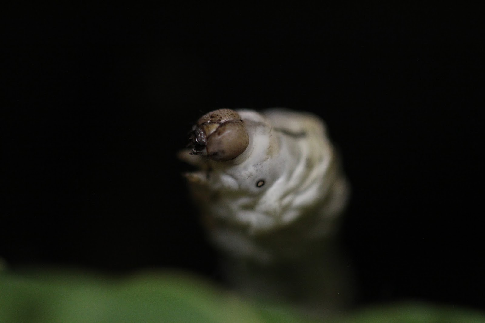
The head consists of six body segments fused together with a cranium. The second, fourth, fifth and sixth segments carry appendages which are modified into antennae, mandibles, maxillae and labium respectively. There are six pairs of ocelli or larval eyes which are located behind and a little above the base of the antennae. There is a pair of antennae formed of five jointed segments and these are used as sensory organs (feelers). The mandibles are well developed and powerful and are adapted for mastication.
In the mouth region the maxillary lobe and maxillary palpi discriminate the taste of food. Also, a median process or spinneret is present through which silk is expelled from the silk gland to form the silk bave or thread. The sensory labial palpi are found on both sides of the spinneret.
(Ref. Textbook of Applied Zoology by P. V. Jabde 2005)


 Photography by M. Vaughan.

Monday, November 2, 2015

Silkworm Photoshoot


They do appear to be posing for the photographer at times!


Photography: M. Vaughan

Moulting - leaving the "old coat" behind!

The skin of the silkworm sheds from the head toward the back, and the silkworm literally "walks out of his skin", leaving it in a little pile behind him. Especially in the later moults while this is happening, the silkworm is "torpid", appearing to be asleep. It's best not to disturb them at this time.

The first moult. 
The skin is receding from behind the head. They still have little black heads.


The second and third moults


Torpid silkworms appear to be sleeping.

Notice the black head has now changed. It came off like a little cap and was left behind.
Skins are left behind in little piles.
The fourth moult.


A skin left behind.


Photography: M. Vaughan

Sunday, November 1, 2015

"Instars" Developmental stages of the silkworm larva/caterpillar


The interval between moults is called an instar. An instar is a stage within a stage. At the end of each instar, the silkworm sheds its skin, enabling growth to continue.  During the shedding of the skin the silkworm appears "asleep".  It stops eating. It is torpid (appearing "frozen" in space) resting on the branch, often with its head slightly raised. It's best not to disturb the silkworm during this time.  After the moult it will happily start munching the mulberry leaves again. The following is an approximate record of the instars of my silkworms.

First Instar  Day 1 to approx. day 4
 After hatching from the egg until the first moult, is the 1st Instar.
The color of the silkworm is blackish and the head is black.

Second Instar  Approx. day 5 to day 11
The period between the first and second moults.


Third Instar Approx. day 13 to day 18
The period between the second and third moults.
 The color of the silkworm is grayish-white. The black head has changed to brown (after leaving the little black "cap" behind. The stripes of the zebra silkworms are showing.


Zebra and the plain silkworms.


Fourth Instar Approx. day 20 to day 26
The period between the third and the fourth moults.


They liked to use the stalks as highways!

Fifth Instar Approx. day 27 to day 35 
The period between the fourth moult and spinning the cocoon.


My silkworms just kept eating and eating!  I understand the silkworm does 80% of its eating during this fifth instar and the silk glands now make up 25% of its body weight.  My largest silkworms were 7cms long, about the size of my little finger. They took their time before starting to spin...seemed too happy munching!


During this lava stage, the silkworm grows from about 3mm at hatching, to about 7cms in length.  It increases it's original size 10,000x by the time it is ready to start spinning its cocoon! Most of this growth is during the fifth instar.

Photography: M. Vaughan.