 |
| Click on photo to enlarge. |
Above is a papered microscope
slide of silkworm larva, scanned directly with a 35mm slide film scanner. Three pairs
of thoracic legs, five pairs of abdominal legs 'pro-legs' and tracheal
structures leading to nine pairs of spiracles (the breathing or respiration pores) are clearly seen. Slide by David Walker, Micscape.
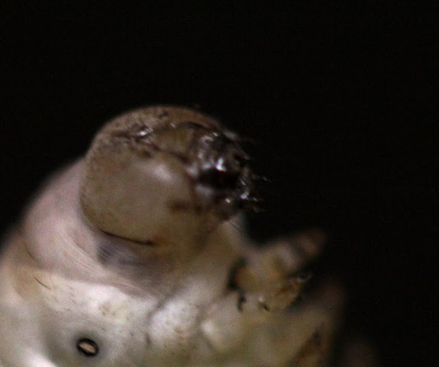
The head consists of six body segments fused together
with a cranium. The second, fourth, fifth and sixth segments carry appendages
which are modified into antennae, mandibles, maxillae and labium respectively.
There are six pairs of ocelli or larval eyes which are located behind and a
little above the base of the antennae. There is a pair of antennae formed of
five jointed segments and these are used as sensory organs (feelers). The
mandibles are well developed and powerful and are adapted for mastication.
In the mouth region the maxillary lobe and maxillary palpi discriminate the taste of food. Also, a median process or spinneret is present through which silk is expelled from the silk gland to form the silk bave or thread. The sensory labial palpi are found on both sides of the spinneret.
(Ref. Textbook of Applied Zoology by P. V. Jabde 2005)
Photography by M. Vaughan.


No comments:
Post a Comment